Oxygen Imaging For Efficient Therapies and Drug Development
Oxygen is a fundamental physiologic parameter with significance to clinical diagnostics, disease treatment, and drug development.
Tissue oxygen maps with our patented technology can incorporate hypoxic and hyperoxic conditions for advancing research in biomedicine.

Frequently Asked Questions
Learn more about our technology and the science behind it.
Oxygen imaging is a technology that generates partial oxygen pressure (pO₂) maps of tissues in vitro and in vivo. O2M's patented oxygen imaging technology is based on pulse electron paramagnetic resonance oxygen imaging (EPROI). EPROI provides fast three-dimensional pO₂ maps non-invasively with high precision and high resolution. Oxygen imaging gives complete oxygen distribution and is distinct from pulse oximetry, polarographic electrodes, or oxygen-quenching luminescence sensors, which provide single point pO₂ values at the location of the probe.
Electron paramagnetic resonance (EPR) is a magnetic resonance technique to detect unpaired electron spins of a molecule placed into a magnetic field by applying radiofrequency electromagnetic field.
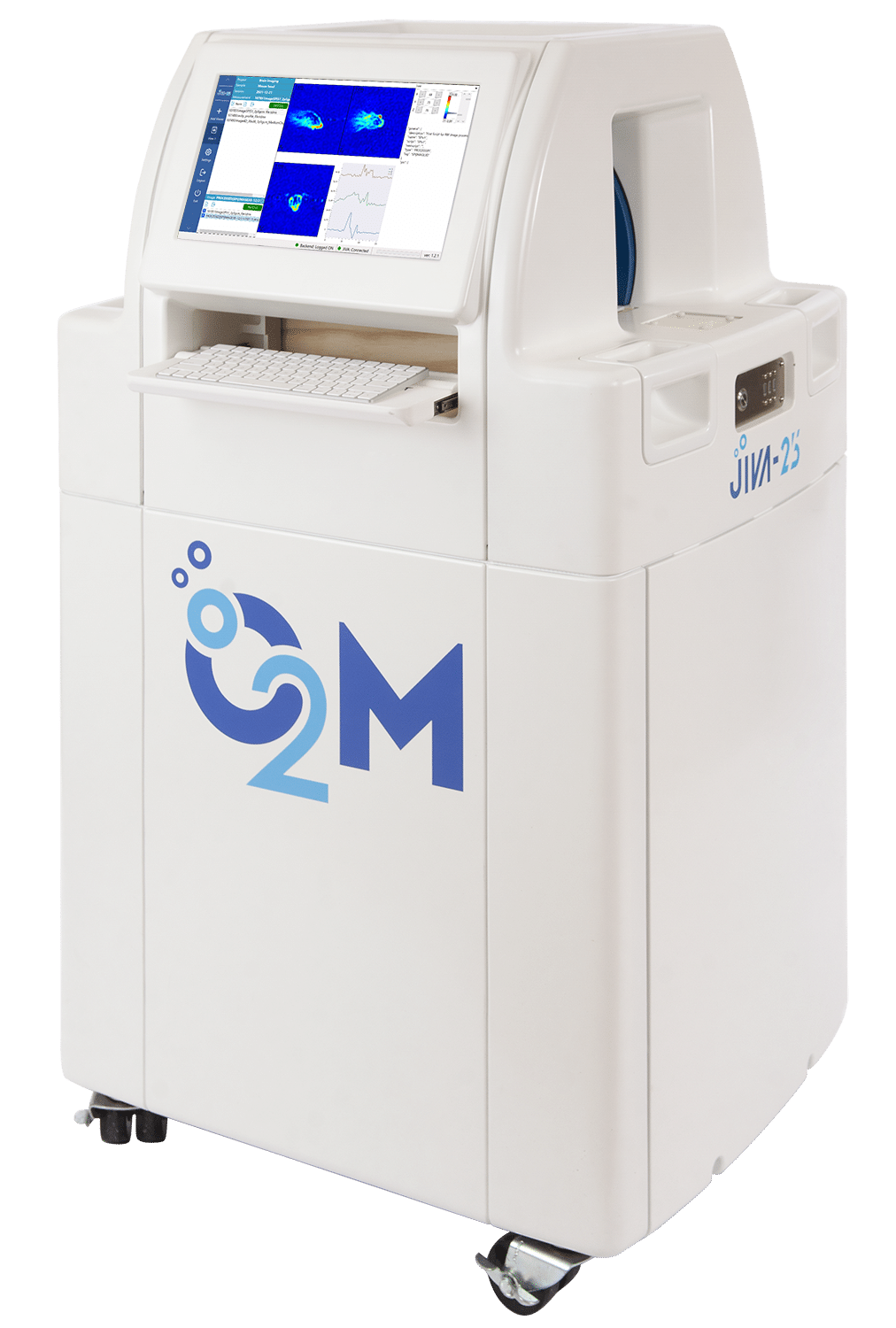
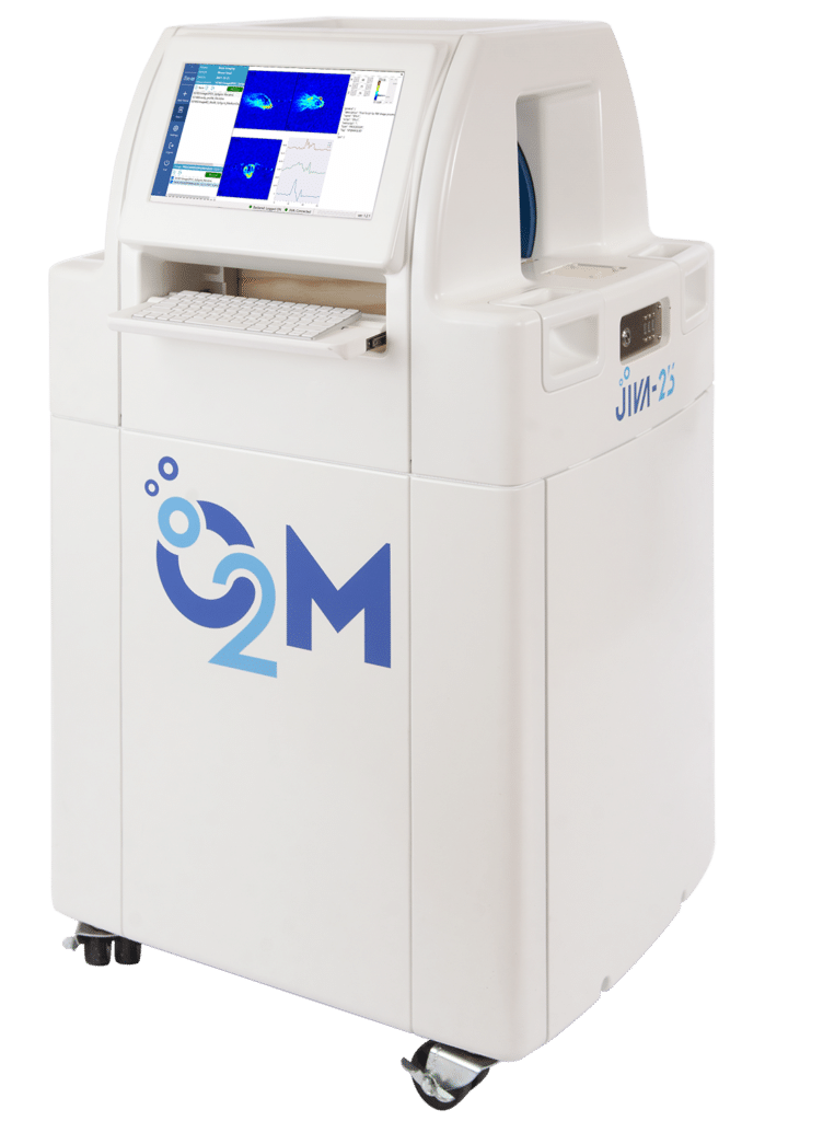
JIVA-25™ is a first-of-its-kind preclinical oxygen imager. JIVA-25™ provides three-dimensional oxygen maps of tissues. Biomaterial samples, cell encapsulation devices, artificial tissue grafts, and small rodents can be imaged with JIVA-25™.
Upcoming Events and Webinars
Our Technology
We use O2M's patented technology to generate 3D oxygen maps.

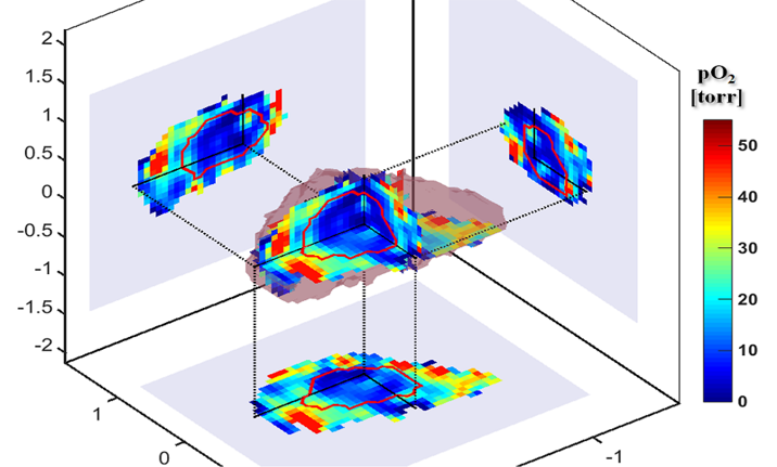
Oxygen Map of a Tumor
Mouse leg bearing a fibrosarcoma. Red line-the tumor margin.
O2M’s core technology, electron paramagnetic resonance oxygen imaging (EPROI), is a non-invasive oxygen mapping method with high precision and absolute accuracy. EPROI detects unpaired electron spins subjected to a constant uniform magnetic field by manipulating them using radio-frequency electromagnetic radiation.
Similar to magnetic resonance imaging (MRI), EPROI uses magnetic field gradients to generate spatial images. In contrast to conventional MRI, EPROI relies on a much smaller magnetic field and constant field gradients. EPROI measures the relaxation maps of a water-soluble oxygen-reporting trityl molecule that distributes in a body upon injection and converts them into oxygen maps.
EPROI applications include but are not limited to:
- Cancer research, diagnostics, and treatment
- Assessment of therapy outcome
- Development of artificial cell replacement devices
- Drug development
- Vitality probing of artificial cell replacement devices
Our Products
Oxygen Imaging for Efficient Therapies.

JIVA-25™
The world's first dedicated preclinical 25 mT oxygen imager. JIVA-25™ provides pO₂ maps with high spatial, temporal, and pO₂ resolution.

CAELI-9™
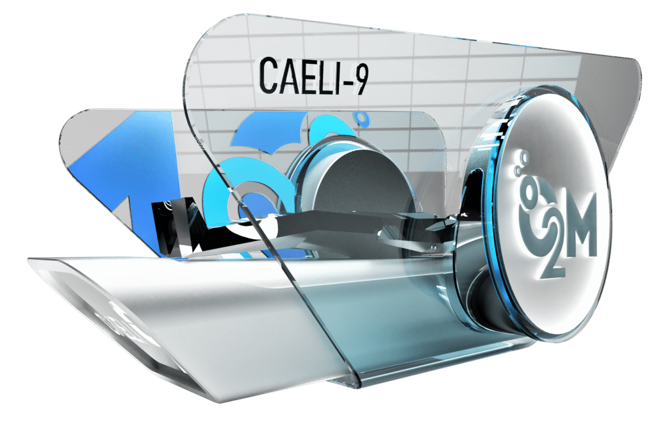
First of its kind human-sized 9 mT oxygen imager. In development, not available for purchase.
Our Services
O2M’s "Oxygen Measurement Core" is a contract research facility. It provides full cycle in vitro and small animal in vivo oxygen measurement services.
All experiments are performed using JIVA-25™. O2M can carry research projects to its full experimental cycle, from sample preparation, cell seeding, and device implantation to in vitro and small animal in vivo oxygen imaging.
O2M can also provide biologic assays and histology assessments, high quality data/images/reports, and support your submission to peer reviewed journals.

In Vivo pO₂ Map (in color) Overlaid on MRI of an Islet Encapsulation Device Implanted Subcutaneously in a C57/BL6 Mouse.
